Why are my shins bowing forward?
Forward bowing of the shins (anterolateral tibial bowing) can result from congenital conditions, nutritional deficiencies like vitamin D, metabolic disorders, or mechanical stress. Medical evaluation including blood tests for bone health markers and imaging studies is essential for proper diagnosis and treatment.


Jump To Section
Understanding Tibial Bowing
If you've noticed that your shins appear to curve or bow forward, you're observing a condition known as tibial bowing or anterolateral bowing of the tibia. This visible curvature of the shin bone can range from a subtle bend to a more pronounced deformity that affects walking and overall leg alignment. While some degree of bowing can be normal during early childhood development, persistent or progressive bowing in adults or older children warrants medical attention.
The tibia, your main shin bone, bears most of your body weight and plays a crucial role in walking, running, and standing. When this bone develops an abnormal forward curve, it can affect not just appearance but also biomechanics, potentially leading to pain, gait abnormalities, and increased stress on other joints. Understanding the underlying causes is essential for proper treatment and prevention of complications.
Common Causes of Forward Shin Bowing
Nutritional Deficiencies
One of the most common and treatable causes of shin bowing is nutritional rickets, primarily caused by vitamin D deficiency. Vitamin D is essential for calcium absorption and bone mineralization. When levels are insufficient, bones become soft and pliable, leading to deformities under the stress of body weight. This condition is particularly common in children but can also affect adults in the form of osteomalacia.

Key Biomarkers for Bone Health Assessment
| Biomarker | Normal Range | Deficiency Level | Clinical Significance | |
|---|---|---|---|---|
| Vitamin D | 25-OH Vitamin D | 30-100 ng/mL | <20 ng/mL | Primary marker for vitamin D status and bone mineralization |
| Calcium | Serum Calcium | 8.5-10.5 mg/dL | <8.5 mg/dL | Essential for bone strength and muscle function |
| Phosphorus | Serum Phosphate | 2.5-4.5 mg/dL | <2.5 mg/dL | Critical for bone mineralization and energy metabolism |
| PTH | Parathyroid Hormone | 10-65 pg/mL | Elevated >65 pg/mL | Regulates calcium and phosphorus balance |
| ALP | Alkaline Phosphatase | 44-147 IU/L | Elevated in rickets | Indicates bone turnover and formation activity |
These biomarkers should be interpreted together for comprehensive bone health assessment.
Beyond vitamin D, deficiencies in calcium, phosphorus, and other minerals can contribute to bone weakness. These nutritional factors are especially important during periods of rapid growth in children and adolescents. Regular monitoring of these essential nutrients through comprehensive blood testing can help identify deficiencies before they lead to structural changes in your bones.
Metabolic and Hormonal Disorders
Several metabolic conditions can affect bone strength and shape. Renal rickets, caused by kidney disease, impairs the body's ability to maintain proper calcium and phosphorus balance. Hypophosphatemic rickets, a genetic condition affecting phosphate metabolism, can cause progressive bowing despite normal vitamin D levels. These conditions often require specialized testing to diagnose properly.
Hormonal imbalances, particularly involving parathyroid hormone (PTH), growth hormone, and thyroid hormones, can also impact bone remodeling and strength. Hyperparathyroidism, for instance, can lead to excessive calcium removal from bones, weakening their structure over time. Understanding your hormone levels through comprehensive testing provides valuable insights into your bone health status.
Congenital and Developmental Conditions
Some individuals are born with or develop tibial bowing due to genetic conditions. Neurofibromatosis type 1 (NF1) is one of the most significant congenital causes of anterolateral tibial bowing. This genetic disorder affects nerve tissue growth and can lead to bone abnormalities, including a characteristic forward and lateral bowing of the tibia that may progress to fracture or pseudarthrosis (false joint formation).
Other developmental conditions include fibrous dysplasia, where normal bone is replaced with fibrous tissue, and osteogenesis imperfecta, a genetic disorder affecting collagen production that results in brittle bones. Congenital pseudarthrosis of the tibia, though rare, represents one of the most challenging orthopedic conditions in children, often requiring multiple surgeries throughout childhood.
Risk Factors and Associated Symptoms
Several factors increase the likelihood of developing tibial bowing. These include inadequate sun exposure, dietary restrictions that limit vitamin D or calcium intake, malabsorption disorders affecting nutrient uptake, chronic kidney or liver disease, and certain medications that interfere with bone metabolism. Additionally, premature infants and those with darker skin living in northern climates face higher risks of vitamin D deficiency.
Symptoms accompanying shin bowing can vary depending on the underlying cause but often include:
- Visible curvature of the legs, particularly noticeable when standing
- Knee or ankle pain due to altered biomechanics
- Difficulty walking or an abnormal gait pattern
- Muscle weakness or cramping in the legs
- Delayed motor milestones in children
- Increased frequency of fractures or stress injuries
- Growth delays or short stature
- Bone pain or tenderness, especially in the shins
Diagnostic Approaches and Testing
Proper diagnosis of tibial bowing requires a comprehensive evaluation combining physical examination, imaging studies, and laboratory tests. Your healthcare provider will assess the degree of bowing, check for associated features like skin changes or muscle weakness, and evaluate your gait pattern. X-rays are typically the first imaging study performed, showing the extent and pattern of bowing while revealing any underlying bone abnormalities.
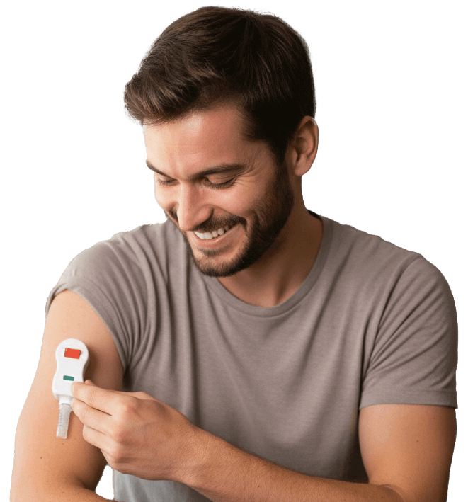
Blood tests play a crucial role in identifying nutritional and metabolic causes of bone deformities. Key biomarkers include vitamin D (25-hydroxyvitamin D), calcium, phosphorus, alkaline phosphatase, and parathyroid hormone. Additional tests might include kidney function markers, thyroid hormones, and specific genetic testing if a hereditary condition is suspected. Regular monitoring of these biomarkers can help track treatment progress and prevent recurrence.
Advanced imaging such as MRI or CT scans may be necessary to evaluate soft tissue involvement, assess bone quality, or plan surgical interventions. In cases of suspected genetic conditions, genetic counseling and testing can provide definitive diagnosis and help predict disease progression. Bone density scans (DEXA) might also be recommended to assess overall bone health and fracture risk.

The Science of Living Well, Longer
Co-authored with Michael Lustgarten, Ph.D. Discover biomarkers that predict healthy aging and add years to your life.
- The critical difference between lifespan and healthspan
- Which biomarkers predict healthy aging at every life stage
- Evidence-based strategies that add years to your life
- How to create your personalized longevity roadmap
Treatment Options and Management Strategies
Medical Management
Treatment for nutritional causes typically involves supplementation with vitamin D, calcium, and other deficient nutrients. The dosage and duration depend on the severity of deficiency and the underlying cause. For vitamin D deficiency rickets, high-dose vitamin D therapy (often 2,000-6,000 IU daily or weekly bolus doses) is prescribed initially, followed by maintenance doses. Calcium supplementation of 500-1,000 mg daily is often recommended alongside vitamin D to optimize bone mineralization.
For metabolic causes, treatment targets the underlying condition. Phosphate supplements and active vitamin D analogs (calcitriol) are used for hypophosphatemic rickets. Kidney disease-related bone problems require careful management of calcium, phosphorus, and vitamin D levels, often with specialized medications like phosphate binders. Hormonal imbalances may need specific treatments such as thyroid hormone replacement or parathyroid surgery.
Orthopedic Interventions
Mild bowing in young children often improves with nutritional treatment alone, but more severe or persistent cases may require orthopedic intervention. Bracing can help guide bone growth in younger children, though its effectiveness varies depending on the underlying cause. Serial casting might be used to gradually correct alignment in flexible deformities.
Surgical options range from guided growth procedures using temporary implants to control bone growth direction, to more complex osteotomies where the bone is cut and realigned. In cases of congenital pseudarthrosis or severe deformity, multiple surgeries throughout childhood may be necessary. External fixation devices, such as the Ilizarov frame, can gradually correct severe deformities while allowing for bone lengthening if needed.
Prevention and Long-term Monitoring
Preventing tibial bowing focuses primarily on maintaining optimal bone health through adequate nutrition and early detection of risk factors. Ensuring sufficient vitamin D intake through diet, supplementation, and appropriate sun exposure is crucial. The recommended daily allowance for vitamin D is 600-800 IU for most adults, though many experts suggest higher amounts for optimal bone health. Calcium intake should be 1,000-1,200 mg daily through diet or supplements.
Regular exercise, particularly weight-bearing activities, helps maintain bone strength and proper alignment. Activities like walking, running, and resistance training stimulate bone remodeling and can help prevent deformities. However, individuals with existing bowing should work with healthcare providers to determine safe exercise levels that won't exacerbate the condition.
For those with treated tibial bowing or at risk for bone deformities, long-term monitoring is essential. This includes regular physical examinations to assess alignment, periodic X-rays to monitor bone structure, and blood tests to ensure nutritional and metabolic parameters remain optimal. Early detection of recurrence or progression allows for timely intervention and better outcomes.
If you're experiencing shin bowing or have concerns about your bone health, understanding your nutritional and metabolic status through comprehensive testing is an important first step. For a detailed analysis of your existing blood test results and personalized recommendations, you can use SiPhox Health's free upload service to get AI-driven insights into your bone health markers and overall wellness profile.
Living with Tibial Bowing: Practical Considerations
Managing daily life with tibial bowing requires attention to both physical and psychological aspects. Physical therapy can help improve gait patterns, strengthen supporting muscles, and reduce pain. Custom orthotics or shoe modifications may compensate for leg length discrepancies or abnormal weight distribution. Swimming and water exercises provide excellent low-impact options for maintaining fitness without stressing the affected bones.
The psychological impact of visible leg deformity, particularly in children and adolescents, shouldn't be overlooked. Support groups, counseling, and connecting with others who have similar conditions can provide valuable emotional support. Early intervention and treatment not only improve physical outcomes but also help maintain self-esteem and quality of life.
For parents of children with tibial bowing, understanding the condition and treatment timeline is crucial. Many cases of nutritional rickets respond well to treatment within months, while congenital conditions may require years of management. Keeping detailed records of growth, treatment responses, and any symptoms helps healthcare providers optimize care over time.
When to Seek Immediate Medical Attention
While gradual changes in leg alignment should be evaluated by a healthcare provider, certain situations require urgent attention. Seek immediate medical care if you experience sudden worsening of bowing, severe pain in the affected leg, signs of fracture such as swelling or inability to bear weight, or skin changes over the bowed area including wounds or color changes. Children showing rapid progression of deformity or failure to meet developmental milestones should be evaluated promptly.
Early intervention often leads to better outcomes, particularly in growing children where bone remodeling potential is highest. Don't hesitate to seek a second opinion if you're concerned about the diagnosis or treatment plan. Specialized pediatric orthopedic centers or metabolic bone disease clinics can provide expertise for complex cases.
References
- Sabharwal, S. (2009). Pediatric lower limb deformities: principles and techniques of management. Springer Science & Business Media.[DOI]
- Munns, C. F., Shaw, N., Kiely, M., et al. (2016). Global consensus recommendations on prevention and management of nutritional rickets. The Journal of Clinical Endocrinology & Metabolism, 101(2), 394-415.[PubMed][DOI]
- Carpenter, T. O., Shaw, N. J., Portale, A. A., et al. (2017). Rickets. Nature Reviews Disease Primers, 3, 17101.[PubMed][DOI]
- Pannier, S., & Glorion, C. (2018). Congenital pseudarthrosis of the tibia. Orthopaedics & Traumatology: Surgery & Research, 104(1), S107-S118.[PubMed][DOI]
- Holick, M. F., Binkley, N. C., Bischoff-Ferrari, H. A., et al. (2011). Evaluation, treatment, and prevention of vitamin D deficiency: an Endocrine Society clinical practice guideline. The Journal of Clinical Endocrinology & Metabolism, 96(7), 1911-1930.[PubMed][DOI]
- Shore, R. M., & Chesney, R. W. (2013). Rickets: Part I. Pediatric Radiology, 43(2), 140-151.[PubMed][DOI]
Was this article helpful?
Frequently Asked Questions
How can I test my vitamin D and bone health markers at home?
What is the difference between tibial bowing and normal leg development in children?
Can tibial bowing be corrected without surgery?
What vitamin D level should I maintain to prevent bone deformities?
How long does it take to see improvement after starting vitamin D treatment?
Is tibial bowing hereditary?
This article is licensed under CC BY 4.0. You are free to share and adapt this material with attribution.


